Category Archives: Services Body

Congenital Hand And Foot Defects

Birth defects of the fingers and toes can occur while the baby is developing in the womb. For example, the baby may have very few fingers or toes. Or she may be born with extra fingers or toes.

In amniotic band syndrome, defects of the hands, fingers, and toes (and other abnormalities) occur when parts of the body become constricted by thin strands of tissue from the amniotic sac. The amniotic sac contains the amniotic fluid that surrounds the developing fetus in the womb. The strands wrap around the fetus, like strings or rubber bands, trapping parts of its body and restricting its growth.

polydactyly

Polydactyly is the presence of extra fingers or toes. The little fingers or the thumbs of the hands and feet are the most frequently duplicated. The extra finger or toe may be just a bulge of fleshy tissue, or it may be fully functional and have its own nerve endings, bones, and joints.


syndactyly

Syndactyly consists of the appearance of an interdigital web between the fingers or toes (the fingers are fused). In syndactyly, the fingers or toes do not spread, resulting in a webbed hand or foot. In simple syndactyly, the soft tissues fuse together. In complex syndactyly, the bones and soft tissues fuse together.

Both polydactyly and syndactyly can occur alone or as part of a genetic syndrome, such as Apert syndrome.

Before birth, doctors can sometimes diagnose these defects during an ultrasound .

After birth, doctors often take x-rays and may do other imaging tests to determine which bones are affected. When the defects appear to run in the child’s family or if doctors suspect that the child has a genetic syndrome, they also examine the child for other physical abnormalities and take a blood sample for genetic testing.

Treatment of finger and toe defects

  • Surgery

  • Prosthesis

Surgery may be done to separate the syndactyly of the fingers or toes and improve function. Surgery may be done to remove an extra finger or toe.

Some children who are missing a finger or toe need a prosthesis, an artificial device that replaces the missing body part.

Congenital Nevus Excision

Congenital Nevus Excision

There are several reasons that patients sometimes decide to undergo the removal of a large nevus. Most of the time, the overriding reasons to remove a congenital pigmented nevus are first to reduce melanoma risk and second to improve appearance which can be fundamental to improving a patient’s overall psychosocial state. Other reasons to remove a nevus may be to improve the strength, texture, sensation, and sweat gland activity of the affected area.

Is Removal Possible?

Small nevi can be removed by simple surgical excision. The nevus is cut out, and the adjacent skin stitched together leaving a small scar. Removal of a large congenital nevus, however, requires replacement of the affected skin. While it is almost impossible to remove every cell of a large nevus, the goal is to remove as many cells as possible while at the same time preserving function and minimizing scarring.

How is Nevus Removal Performed?

When small nevi are removed, the surrounding skin is simply pulled together and stitched up. Removal of a large nevus involves the replacement of the affected skin. Skin can be transferred from another area of the body (grafting), or adjacent skin can be stretched, then used to cover the area where the nevus was removed (tissue expansion). There are many details about the surgery you should consider and it is best to consult a surgeon experienced in the treatment of giant nevi.

There are limits to skin grafting and tissue expansion procedures. You can only harvest so much additional skin from one person. Can another person donate skin? Not as easily as you might think. This is not commonly done and presents many risks and challenges. Can’t they just grow some artificial skin like I saw on TV? No, sorry. That skin does not have all the other tissue underneath it that the patient needs like muscle, fat and blood vessels. Artificial skin can keep a burn victim alive, but it is usually not a viable option for nevus removal.

Hemangioma

Infantile hemangioma or childhood hemangioma is a benign vascular tumor caused by abnormal growth of blood vessels that do not offer risk to the health of the individual, however, may disturb by appearance and location.

Hemangioma is more common in newborn girls and may be single or multiple lesions located on the face, scalp, or trunk.

How to Identify Hemangioma?

It is possible to identify the superficial lesion with red and bright color, similar to the color of the strawberry shortly after birth. When they are deeper lesions, they have bluish or violet tint. During the first year of life, hemangioma has rapid growth in volume extension, especially in the first six months of the child’s life.

From this period, the growth of the lesion is interrupted and the involution process may occur, which allows the total disappearance of the condition throughout life without the need for medication or surgery.

However, some cases of hemangioma may be more complicated, such as ulceration, especially in lesions located in areas of friction such as accommodation regions of diapers, elbows, shoulders, knees and lips. Frictional ulceration can cause pain, bleeding, and secondary infections. In cases of hemangioma in the eye area, the lesion may compromise vision, especially those located on the eyelids.

Children who have more than five lesions in the body should undergo imaging tests, such as ultrasound, tomography or RESONANCE for the identification of hemangioma in the internal organs, especially in the liver, a condition that does not offer health risk and may disappear over time.

Hemangiomas That Are Indicative of Treatment:

There is not yet a standard protocol for the treatment of hemangiomas, but in specific situations treatment should be initiated in the following cases:

  • 1.Palpebral occlusion – eyelid occlusion in the first months of life can cause amaurosis due to lack of luminous stimulus or amblyopia.
  • 2.Genital/Anal – often grow and ulcerate causing a painful ulcer, in addition to tissue loss and aesthetic disfigurement – In addition, they are generally complicated by secondary infection.
  • 3.Labial/Palpebral Edge and Auditory Conductive (Periorificial) – often ulcerate causing aesthetic disfigurement.
  • 4.Nose tip – grow a lot in volume and, secondary form, can promote the destruction of the nose cartilages. It also produces aesthetic disfigurement. They tend to regress very slowly and leave a permanent nasal deformity, difficult to correct.
  • 5.Extensive Segmentals – Phace Syndrome, Pelvis and Sacral.
  • 6. – Any location with accelerated growth and significant aesthetic impairment.
  • 7.Congenital Nich – This hemangioma may require treatment depending on location. The tendency is to grow and not regress over time.
  • 8.Kasabach-Merrit syndrome – Most cases have histology of kaposiform hemangioendothelioma or tufted hemangioma. The lesion increases in volume, leading to thrombocytopenia (platelet hijacker hemangioma), CIVD and eventually death.
  • 9.Visceral (Multiple Hemangiomatosis with Visceral Involvement) – hemangiomas may be present in the liver and CNS.

How to Treat Hemangioma

In general, after the diagnosis of hemangioma, specialists seek to perform early treatment to reduce growth and the chances of sequelae that may compromise the child’s aesthetic health. In general, after the diagnosis of hemangioma, specialists seek to perform early treatment to reduce growth and the chances of sequelae that may compromise the child’s aesthetic health. The treatment indicated for small lesions consists in the use of beta-blocker medications (propanalol), used to treat heart problems and was accidentally discovered for its action that prevents the growth of hemangioma. The drug should be used in accordance with medical guidelines.

Hemangioma Surgery

Hemangioma surgery is recommended in cases of extensive injuries that hinder movement or compromise the aesthetic health of the patient. The procedure consists of removal of hemangioma and marriage to healthy skin.

Hemangioma surgery is considered a simple procedure that offers low risk of life.

How is the Postoperative

The postoperative surgery of hemangioma is considered simple and offers little care that should be followed according to the guidelines of the plastic surgeon:

  • The use of analgesics should be done according to the dose given by the specialist to eliminate pain and discomfort in the first days after the procedure. In cases of severe pain accompanied by fever, the doctor should be sought immediately.
  • It is recommended that you avoid exposure to the sun for 30 days to prevent the formation of stains at the healing site.

How to Do Hemangioma Surgery?

Dr Vishal Patel is a plastic surgeon with specialization in child plastic surgery and hemangioma surgery. Contact us and make an assessment to take care of your child’s health and quality of life.

Leg Defects

Lower extremity reconstruction is an essential part of plastic surgery and focuses on the treatment of injuries and defects caused by trauma, cancer, or chronic disease processes. Over the past 25 years, advances in plastic surgery techniques such as free tissue transfer and more advanced wound care technologies have revolutionized the field, allowing limbs that would otherwise have been amputated to be saved.

The goal of lower extremity reconstruction is to protect/cover open leg defects and wounds to heal the patient’s wound and allow them to resume life, mobility, and return to work, while simultaneously avoiding amputation. Defects and open wounds in the lower extremity are caused by trauma, tumor resection, and chronic diseases such as peripheral vascular disease and diabetes; these wounds need reconstruction for several reasons.

First, any exposed bone that is not covered by vascularized soft tissue is at risk for osteomyelitis, bone necrosis, and sepsis. Osteomyelitis is mostly due to amputation in patients after leg trauma, or in patients with systemic diseases, mainly diabetes. Second, open wounds lead to chronic pain, inability to move freely, significant medical bills, and unemployment. Exposed tendons begin to dry out and become necrotic, and exposed blood vessels are at risk of rupturing.

Diabetic Foot

Plastic surgery allows to address the foot of diabetics preventively or to correct the complications of the disease. Thanks to the reconstruction of ulcers it is possible to avoid amputations and we will explain why.

Plastic surgery and diabetic foot

The appearance of ulcers on the feet of a diabetic patient is the result of a nerve injury called diabetic neuropathy. Consequently, the nerves are affected in their three components. On the one hand, at the sensory level, since there are alterations in the sensitivity of the foot, being unprotected and more loaded.

On the other hand, diabetic neuropathy produces alterations in the form of support of planting, which causes you to lose the normal way of walking. At the autonomous level, it is responsible for the appearance of dryness on the skin.

To prevent the appearance of diabetic foot, daily monitoring is necessary to prevent the development of these lesions. It is necessary to recognize existing deformities in order to treat them before ulcers form.

There are several ways to apply plastic surgery to diabetic foot. First, preventively, this technique can perform posterior tibial nerve desiccation to improve neuropathy in case patients have lost sensation.

On the other hand, it is important for the reconstruction of foot ulcers to avoid amputations. This requires a vascular and traumatological study, in addition to ruling out that there are infections in the bone. When there are healthy vessels and there is no infection, we can consider reconstruction so as not to amputate.

To proceed with the reconstruction, transplants of healthy and vascularized tissues are carried out from other locations of the body such as the thigh, which join arteries and veins of the feet. Without exquisite care of the intervened foot and proper control of the disease, ulcers will come out again in a short time.

Given the challenges of healing complicated wounds in patients with diabetes, use of plastic surgery techniques ranging from partial thickness skin grafts and local random flaps to muscle flaps and pedicles is essential.

The global impact of diabetic foot has made very clear the need for robust and durable surgical options to facilitate rapid closure of wounds.

Treatment of diabetic foot wound challenges the surgeon with multiple obstacles on the way to eventual wound closure. There are numerous options available and one must be familiar with each modality in order to select the best treatment for the patient. Surgeons must also ensure a thorough diagnosis and carefully consider a number of factors (patient age, vascular status, comorbidities, adherence, etc.) to determine the best course of treatment.

Plastic surgery techniques for soft tissue reconstruction have proven successful for long-term healing and stabilization. Accelerated healing of these complicated wounds not only improves the patient’s quality of life, but can also significantly reduce the healthcare costs associated with prolonged wound care. These techniques require meticulous perioperative planning.

The fundamental goals of plastic surgery techniques in wound closure have not changed since ancient times: preservation of form and function. A deep understanding of the gradual approach to the use of such procedures is imperative to obtain successful results.

Regardless of the approach you use, the main goal is to achieve a healthy, uninfected wound with adequate perfusion and biomechanical stability. Ultimately, you strive to achieve a plantigrade and functional foot without residual and recurrent ulcerative lesions.

Surgeons can perform concomitant bone reconstructive procedures to correct the underlying deformity and further promote foot stabilization for soft tissue reconstruction. In addition, proper discharge techniques, such as a complete cast, ferulization or external fixation with assistive devices, are vital to the durability of soft tissue reconstruction of soft tissue reconstruction.

bedsore treatment in Mumbai

Bedsore Management

A common problem in older adults with reduced mobility or in patients with long periods of rest is skin lesions. These are produced by pressure and friction of the support surfaces and are called bedsores or decubitus lesions or pressure ulcers.

Areas of the body around protruding bones tend to be more prone to bedsores than areas with more fat. In their task of healing bedsores at home, nurses observe that bedsores most often form in:

  • Coccyx or buttocks
  • Scapulae and spine
  • Back of the arms and legs (where they rest in contact with the chair, for example)
  • Back or sides of the head
  • Shoulder blades
  • Heels, ankles and skin behind the knees

People who are most at risk for bedsores have medical conditions that limit their ability to change positions or cause them to spend most of their time in a bed or chair.

Why do bedsores form?

Bedsores are caused by 3 main factors:

  • By pressure: immobility causes constant pressure in an area of the body and prevents blood from circulating. The tissues do not receive oxygen and nutrients and can die in that area, causing the injury.
  • Friction:skin contact with bedding or the patient, or with diapers, especially if the skin is moist, can cause this injury.
  • By friction: rubbing against a surface, for example, when sliding on the bed or from the bed to the chair, the skin can be injured in vulnerable areas.

Bedsores can develop over the course of hours or days. Most lesions heal with treatment, but some never heal completely and require surgical treatments, PRP infiltration, or the use of special dressings.

Warning signs of bedsores

  1. Unusual changes in skin color or texture.
  2. Edema in a support area on bone.
  3. Drainage initially serohematic and, as the days go by, purulent in appearance.
  4. An area of skin that feels colder or warmer to the touch than other areas or with increased sensitivity.
  5. Pressure ulcers are differentiated by their depth, severity, and other characteristics.
  6. The degree of skin and tissue damage ranges from red, intact skin to a deep injury that affects muscles and bones.

bedsore treatment in Mumbai
Treatments

The treatment of bedsores depends on the depth of the affected tissue, and they are classified in levels from 1 to 4, with level 1 being that of the most superficial lesion.

A bedsore is unlikely to heal quickly. Even when there is home healing treatment, it can take several weeks, due to damage to the skin and other tissues.

Less severe pressure ulcers often heal within a few weeks with proper treatment, but serious wounds may need surgery.

In stages 1 and 2, when the epidermis and part of the dermis are affected, prolonged treatment is generally effective, and the bedsore heals. At levels 3 and 4, when deep dermis, muscle and even bone are already affected, the bedsores become infected and can be fatal. There is a treatment option with cleaning by surgery.

So, the importance of daily skin inspection is precisely to find the vulnerable areas reddened before the bedsore forms. If it has become formed, the call to the doctor and the start of treatment should not exceed 24 hours.

Home care services may involve more than one professional, such as a GP, plastic surgeon, nutritionist, physiotherapist or nurse. They will indicate what the treatment should be and what to expect from it. If the patient does not have a professional nurse 24 hours a day, which is the most frequent, the doctor will indicate the home service, one or more times a day. It will also give the necessary indications to the caregiver or family companion. Indications may vary, but generally include:

  • Repositioning: avoid contact of the bedsore with the surfaces of the bed or chair.
  • Use support surfaces, such as cushions, that help you sit or lie down in another way.
  • Cleaning and drying of the wound, with indicated products.
  • Bandage to create a barrier against infections. Bandages may have impregnated creams or medications.
  • Removal of damaged or dead tissue.
  • Medications to control pain.
  • Medications to prevent or stop infections.
  • Diet rich in protein.
  • Surgery and graft if it’s a deep bedsore or if the previous treatment has failed.

There are other supportive therapies, such as the application of negative pressure, performed by a physiotherapist in the affected area, to remove damaged tissues and induce blood circulation. Also, hyperbaric pressure therapy or growth factors. They are optional treatments that are combined with the previous ones.

Preventive Measures

The first recommendations are the change of position frequently to avoid stress on the skin, the maintenance of good nutrition and fluid intake, the cessation of smoking and promote passive mobility at regular intervals of the bedridden patient.

Therefore you should change the weight support point frequently (1 time per hour), make the patient walk if possible, or use a special wheelchair (some wheelchairs may tilt, and this can relieve pressure), use cushions or a special pneumatic mattress to relieve pressure and ensure that the body is well positioned and adjust the elevation of the bed (if the bed can be raised in the head part, raise it no more than 30 degrees) to help prevent the patient from slipping.

Also consider the following skin care suggestions: keeping skin clean and dry with a gentle cleanser to limit skin exposure to moisture, urine, and feces; protect the skin with moisture barrier creams to protect the skin from urine and feces; change bedding and clothing frequently by paying attention to clothing buttons and wrinkles on sheets that can irritate the skin. Remember to check the skin daily by watching carefully for warning signs of a pressure sore.

Repair of Tendon/Nerve/Artery

The Peripheral Nerve is responsible for taking the commands from the brain to the muscles so that they perform movements, in addition to bringing the stimuli of pain, sensitivity, pressure and touch from the periphery to the brain, so that they can be interpreted. After a cut on the hand, the patient should immediately seek a hospital. Every cut on the hand must be urgently examined by a trained doctor.

What is a Peripheral Nerve?

The nerve is an extremely delicate structure that is shaped like a tube and has the main function of carrying nerve stimuli (electrical currents) from the brain to the extremities of the limbs and bringing them back. They work like a light cable, carrying and bringing energy and commands, which will be interpreted by the different organs.

The nerve functions as a large hollow cable filled with extensions of nerve cells. The processes that carry stimuli from the brain to the periphery are axons and those that bring them back are dendrites.

What is the Function of a Peripheral Nerve?

The nerve is responsible for taking the commands from the brain to the muscles so that they perform movements, in addition to bringing the stimuli of pain, sensitivity, pressure and touch from the periphery to the brain, so that they can be interpreted.

Nerves also carry the command for the correct functioning of blood vessels (arteries and veins), controlling their caliber and blood flow, in addition to controlling the function and growth of hair, skin and nails.

What Care Should You Take After a Cut?

Before going to a hospital, clean the wound and cover it with a clean cloth or towel. A tourniquet, belt or tourniquet should not be placed to reduce local circulation. This only increases tissue damage. When there is considerable bleeding, a compressive dressing can be applied to the wound. This pressure will stop bleeding. Also, no bleeding vessel should be clamped with forceps. Clamping will make surgical reconstruction impossible.

After a cut on the hand, the patient should immediately seek a hospital. Every cut on the hand must be urgently examined by a trained doctor. An open wound, including only the skin, has a fair amount of time to close. Generally, a cut is not sutured after six hours of evolution, as the risk of infection increases.

After an accident, the patient must stop eating and drinking. In case of urgent surgery, anesthesia will be safer if the patient is fasting.

Whenever it is suspected that a cut has caused a nerve rupture, the patient should not be anesthetized before all tests have been carried out. The infiltration of Xylocaine in the wound will cause the nerve to lose its function for a period of up to 10 hours, making it impossible to define the diagnosis.

What Does the Patient Feel With a Nerve Injury?

Initially, the patient feels loss of skin sensitivity in the region distal to the cut. She will be anesthetized there. Often the patient remains with protective sensitivity. That is, he even feels that some object causes an injury or burn to the skin, but he cannot determine precisely what it is.

He will also lose control of the muscles that are controlled by that nerve. Muscles become flaccid and functionless, which can lead to joint deformities.

Whenever there is a cut at the anatomical location of a nerve’s path and there is numbness in the skin, there is a strong suspicion of a nerve rupture. However, there are different types of injuries, which can be partial and total.

How is the Diagnosis Made?

The diagnosis of nerve damage is clinical, made by the hand surgeon when examining the patient. To determine the severity of the injury (to know if it was complete), there are specific tests.

One of them is called two-point discrimination. With normal fingertip sensitivity it is possible to differentiate the touch of two sharp objects less than 5 millimeters apart. When there is a cut and the patient cannot distinguish two points that are more than 10 millimeters apart, it is known that the nerve is completely ruptured.

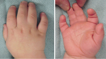
Another test that can be used, especially for pediatric or difficult-to-cooperate patients, is to place the extremity in a basin of water. Normally innervated skin becomes wrinkled when exposed to water for a long time. If the innervation of this region is damaged, the skin remains smooth.

At the site of the cut, whenever there is damage to a nerve, there will be a sensation of shock when being touched, similar to a bare electrical wire. We call this the Tinel Signal.

Tests used for chronic and compressive nerve injuries, such as Electroneuromyography, are not very useful in acute trauma. The results are often confused due to the passage of some electrical discharge to the wound site and the overlapping innervation in the skin.

Is Every Peripheral Nerve Injury Treated With Surgery?

First, it is important to understand the anatomical location of the nerve. Every injury that affects the body of the neuron does not have complete recovery. Neuron bodies are located in the brain (head) or spinal cord (spine). Therefore, injuries in these places, in the vast majority of cases, leave serious, often irreversible, sequelae. On the other hand, lesions that affect the extensions of neurons (peripheral nerves) are capable of regeneration (growth). But for this recovery to be effective and organized, the external membrane that covers the nerve must be intact.

It is also important to differentiate the types of injuries. Injury that results from an open wound, such as a cut, crush, or gunshot wound, should be treated with surgery. Closed lesions, in which there is no opening in the skin, usually do not need surgery.

What is Peripheral Nerve Surgery?

In surgery, the two ends of the nerve are connected, so that the extensions of the neurons can grow in an organized way. This is an extremely delicate surgery, as the nerves are often a millimeter in diameter or less.

After a cut, the nerve works like a rubber band, that is, it shortens over time. The aim of the surgery is to remake the protective membrane of the nerve and align the extensions of the neurons, thus preventing the two parts of the tube from becoming separated.

Due to the small caliber and delicacy of the nerves, for its reconstruction we use materials and instruments made specifically for this type of surgery. They are called microsurgical materials. They are microscopes, magnifying glasses, tweezers, scissors and suture threads (these are often thinner than hairs).

How is Recovery?

The extensions of neurons have the ability to regenerate and grow after injury. Thus, whenever there is a cut in a peripheral nerve, there is an attempt to grow it back to its original location.

Once injured, the peripheral nerve will enter a period of rest for 30 days. During this period it does not grow or regenerate. After that, the extensions of the neurons will grow by an average of one millimeter per day. It will be 3 centimeters per month. Therefore, the further the injury occurs in relation to the target organ, the longer the recovery.

This recovery can be followed through the Tinel Sign. Progressively, the patient begins to feel the shock in a position further away from the injury site.

What is a Neuroma?

Neuroma is a kind of tumor that forms in the disorganized attempt of nerve growth and regeneration. It occurs when the nerve grows and does not “find” the other side of the tube (the two ends of the nerve are separated). One end becomes bulky and extremely sensitive by exposing neurons without a protective layer.

This painful swelling of the nerve tip is called a neuroma.

What are the Factors that influence the Treatment?

There are factors related to the patient, the surgery and the type of injury.

The younger the patient, the greater the ability of the nerve to regenerate, especially in children. In adults, less regeneration is observed after age 60. A clean cut, by knife or guillotine, is easier to treat when compared to crushing or pulling injuries. Systemic diseases, such as diabetes, also have a detrimental effect on regeneration. A long period between cutting and surgery is also harmful. The location of the cut also influences. Cuts on the fingers heal faster than cuts on the elbow or arm.

Changes in other tissues associated with nerve injuries, such as fractures, skin loss, or tendon injury, also alter the nerve’s recovery. Each tissue has a specific treatment and a period of regeneration and rest. So when there are several injuries at the same time, one can interfere with the healing of the other.

Another factor that we have to avoid and that has a negative effect on treatment is infection.

How is a chronic injury treated?

Any injury that is not treated in the initial phase is more difficult. Nerve damage is considered chronic after three weeks of evolution. In the first few days after the trauma, the surgeon can easily bring the ends of the nerve cables to their original position and connect them. However, after a few weeks, the ends shorten and it is no longer possible to place them against each other.

For these cases, we use a nerve graft. Nerves are taken from another location and function as bridges connecting the two ends. They are a kind of tube that allows the growth of the extensions of neurons inside.

The nerves used as a graft are less important than the injured one and their removal will not cause functional impairment. The most used is the Sural Nerve, from the lateral portion of the leg. It is only sensitive and is relatively thick and long. It is responsible for the sensitivity of a small lateral portion of the foot, and its removal is well tolerated.

Whenever possible, we use nerves from the same limb as the lesion as a graft. In the arm we used the Medial Cutaneous Nerve of the Forearm. It has similar characteristics to the Sural Nerve, being responsible for the sensitivity of the inner portion of the forearm.

There is no time limit for the recovery of sensory nerves, even after years they can be reconstructed. The motor nerves (which control muscles) must be treated within a year, after which there is a definite atrophy of the muscles. For these patients and also for cases where there is not a complete recovery after the surgery, we can do tendon transfers. They consist of using functioning tendons and muscles to replace paralyzed ones.

How is it after surgery?

It is important for the patient to be aware that recovery is slow, and can take up to a year. Often, after surgery, no change in symptoms is noticed. Improvement will only be seen weeks or months later, with the growth of the nerve and the return of sensitivity and muscle function.

Symptoms such as shock at the wound site or a burning sensation in the skin may improve soon after surgery.

Is Physiotherapy Necessary After Surgery?

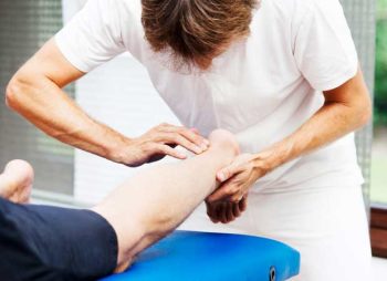
Rehabilitation after peripheral nerve surgery is primarily aimed at restoring the function of tissues that were not injured in the trauma, such as muscles, tendons and joints. Immobilization of these structures causes atrophy and stiffness that is difficult to recover.

Nerve regeneration takes a long time, but there are activities and exercises that encourage this recovery. The use of different stimuli on the anesthetized part makes the nerve recover faster.

What are the Changes for the Future?

Research carried out mainly on laboratory animals shows that certain substances and cells have the ability to stimulate faster nerve growth. Among them are growth factors, stem cells and platelet-rich plasma. These substances are present in the patient and are removed and concentrated before being used.

There have also been attempts to improve nerve grafts. Artificial tubes (absorbable or not) and natural tubes (such as veins and arteries) were tested trying to avoid a second incision for the removal of the nerve graft. Several studies have shown good results, but nothing has been superior to the classic nerve graft procedure.

Thigh Lift

The thigh lift is a surgical procedure that removes excess skin and fat from the inside or outside of the thigh. This surgery is usually a desirable procedure after a large weight loss and when diet or exercise fail to firm loose skin. Liposuction can be used as a fit on the contour of the inner thigh. If you are committed to maintaining a healthy lifestyle and a moderate weight, thigh lifting can help you regain the contours of the thigh you want.

What is the thigh lift surgery?

The thigh lift surgery is an operation that surgically removes excess skin and adipose tissue to recover sagging in the inner-outer region of the thigh and hip. It can be performed simultaneously with the body lifting operation (lipectomy) to improve the overall contours of the body.
Different methods can be used in the thigh lift surgery. Depending on the degree of need of the body, lipolysis laser, lipomatic or liposuction are some of them.
Leg stretching procedures, used in cases where applications such as sports, diet and massage are insufficient, recover relaxation in the thigh region with soft tissues and aim to correct deformations.

Advantages of thigh lift

Leg stretching surgery, required in cases such as sudden excessive weight loss, post-pregnancy, is the easiest and fastest process of recovery from sagging observed with loss of elasticity. It is the only way to treat excessive sagging that cannot be solved with sports or a healthy diet.
Sagging in the thigh region can sometimes be at a level that can cause discomfort in patients due to friction. Especially in summer, this friction brings problems such as rash or fungi, depending on the clothing used. Considering all these problems, thigh stretching becomes necessary. As this procedure avoids health problems and also eliminates the aesthetic concerns of the person, it is recommended that people with sagging thigh consult a specialist.

What should be considered before surgery?

Since it affects the blood and oxygen going into the tissues, smoking should be stopped 2 to 3 months before the operation. The person should also stop using anticoagulants in the 2 weeks prior to surgery. If there is a medical condition, such as high blood pressure, it should be checked before surgery. When examining a series of tests requested by the expert, the operation process can be decided when no problems are seen.

Who can be done?

The stretching process, in which excess tissues of the leg skin are removed and tightened in the desired shape, can be applied to anyone over 18 who requires this procedure. It is preferable in loosening the skin by excessive weight loss, excess in the leg region after birth, sagging of the skin of the leg with age. In examinations performed by specialists of the promed clinic, the process is planned if there is no obstacle to surgery.

A good candidate for thigh lifting

  1. Who has a significant looseness on the skin of the arm
  2. Who has relatively stable weight and who is not significantly overweight
  3. Healthy
  4. Non-smokers
  5. Individuals with a positive outlook and realistic expectations.

Preparation for thigh lifting surgery

  1. Consult the surgeon for your goals, perform a physical examination, a laboratory test, chest x-ray and a medical evaluation
  2. Take certain medications or adjust your current medications as instructed by your surgeon
  3. Stop smoking before and after surgery two weeks
  4. Avoid taking aspirin, anti-inflammatory drugs and herbal supplements as they may increase bleeding

Thigh lifting procedure:

  1. Surgery is performed under general anesthesia.
  2. The surgeon marks the incisions to remove excess skin and fat. Depending on the desired amount, the incision may extend around the thigh or only part of the path around the inner part of the thigh.
  3. After the incision, the surgeon elevates the skin and fat of the underlying muscles of the thighs and removes excess skin and fat to reshape the thighs. Drains can be placed under the incision.
  4. The incisions are carefully closed in several layers of strong sutures to minimize their scarring. The surgeon uses a technique in which the sutures are under the skin and are absorbed by the body. The skin will be sewn.

The lifting surgery of the thigh takes approximately 4-5 hours.

Postoperative care for thigh lifting

  • Wear compression clothing to support the thighs and decrease swelling for at least 6-8 weeks
  • During healing, it is necessary to lift the knee when sleeping to reduce swelling.
  • Typical symptoms after surgery are bruising, swelling and some pain within a few weeks. Contact the surgeon if the pain, redness and swelling are signs of a problem.
  • Remove the stitches after surgery seven days.
  • Attend from all medical appointments.
  • Avoid strenuous activities that interfere with the thighs to prevent scar rupture. At least for six weeks.

Risks and complications:

  • Scarring
  • The scar is quite long and dressings are needed every day to prevent infections.
  • Patients should wear compression clothing, required 24 hours a day, for at least one month. The compression piece helps support the legs during healing, decreasing postoperative swelling and preventing the scar.
  • Bad healing, maybe ruptured. The suture will occur again.
  • Fluid accumulation (seroma)
  • Damage to deeper structures such as nerves, blood vessels and muscles
  • Necrosis of fat and tissue.
  • Numbness or decreased skin sensation
  • Pain
  • Asymmetry

Thigh lifting recovery:

A sterile dressing is applied to incisions every day and a compression garment is worn on the thighs for at least a month. This compression piece helps support the legs during healing, decreases postoperative swelling and helps reduce any bruising that may occur. You can return to work after surgery a week, but should avoid strenuous activities for six weeks. The scars will be visible, but they could disappear for a year.

arm lift surgery in Mumbai

Arms Reshaping

Brachioplasty (commonly called arm lift surgery in Mumbai) is a surgical procedure used to reshape a person’s arm to meet the aesthetic goals of their body.

Physical exercises can improve the muscle tone of the arms, but they do not always solve the problem of localized fat, much less excess skin in this region. Lipodystrophy and flaccidity of the arms affect self-esteem and generate discomfort. People with these alterations can feel embarrassed when wearing dug clothes or even the simple act of saying goodbye, moving the arm.

Arms reshaping surgery in Mumbai targets the lower arm (or triceps) from the top of the arm down to the elbow. Excess skin is removed from the arm, remodeling the area to give the arm more tone and definition. The result is a more proportional and toned arm.

What is arm lifting surgery in Mumbai?

An arm lift, or brachyplasty in Mumbai, is a surgical procedure that:

  • Reduces excess sagging skin that leans down
  • Tightens and smoothes the underlying supporting tissue that defines the shape of the arm
  • Reduces localized pockets of fat in the arm region

Weight fluctuations, aging and heredity can cause the upper arms to have a drooping and flaccid appearance. This is a condition that cannot be corrected through exercise.

Arm lifting surgery can be right for you if the lower part of your arms are flaccid or appear loose and full due to excess skin and fat.

Why look for arm plastic surgery?

There are many factors that contribute to loose and flaccid arms. Some of the most common reasons for arm plastic surgery are:

  • Weight fluctuations, including significant weight loss.
  • Loss of elasticity in the skin due to age.
  • Weakened muscles or tissues.
  • Lack of definition.

Exercise can sometimes help sagging the skin under the arms, but often not enough, especially after a significant weight loss. By surgically removing excess skin and remodeling the arm, a…

… Person can have their arms in good condition and sided that they have ever had or have always wanted.

Brachioplasty in Mumbai

Surgical procedure of arm plastic

A general procedure of remodeling the arm lasts between one to three hours and can be performed with general anesthesia or anesthesia at dusk (a form of anesthesia that leaves you awake but unaware of the procedure). Liposuction in Mumbai may be included in the procedure, when necessary, to remove any localized fat deposits that impair the final results of arm lifting surgery. All incisions are placed under the arm to hide and/or minimize scars.

Although there is some downtime associated with recovery for an arm lifting surgery in Mumbai, most people are performing light activities within a few weeks and usually return to the normal routine, including lifting, within six weeks.

Arm lifting techniques

An incision is made inside the arm, above the elbow to the armpit. Excess skin is removed and, if necessary, fat is aspirated from the arm. Depending on the degree of excess skin, the incision can be made from the elbow to the armpit, or just inside the armpit.

Anaesthesia

Brachioplasty in Mumbai is performed under local or general anesthesia, depending on the length of the procedure.

Before surgery

  • Avoid taking aspirin or other products containing aspirin for two weeks.
  • If you are a smoker, stop smoking to help with healing.

Recovery after arm lifting

  • Most patients resume normal activity in 1-2 weeks.
  • Full recovery takes several weeks.
  • Depending on the extent of excess skin, the scar may be limited to the armpit, or may extend inside the arm, from the top of the elbow to the armpit.

Is there a scar after arm lifting?

The operation is performed by means of a thin incision from the inside of the elbow to the armpit. After the stitches are removed, a dressing is made in the hospital once a week. As it is usually an invisible area, the suture mark here is not very noticeable. Although the scars do not completely disappear, their prominence will decrease in 2 months.

Post-operation healing process

The recovery period after arm lifting surgery in Mumbai is shorter when local anesthesia is applied compared to other surgeries. On the recommendation of the specialist physician, general anesthesia can also be applied in some cases of excessive sagging, in these cases the recovery time may also be affected. The person is invited to stay in the hospital for 1 day.

It is recommended by plastic surgeon in Mumbai to use a corset for the arm for the period recommended by the specialist doctor after surgery. It is an application that helps the subcutaneous tissue recover more quickly. In addition, in possible pain situations, analgesics can be used on the recommendation of the doctor. The average recovery time is 2-3 weeks. Those who do arm lifting surgery in Mumbai should avoid heavy work during this process.

Mommy Makeover Surgery

Mommy Makeover refers to a combination of procedures in a single surgical act, relating to breast plastic surgery and abdominal plastic surgery. It is a surgical intervention to improve the contour of the breast and abdomen, which are often altered after pregnancy.

After one or more pregnancies and the breastfeeding period, women undergo significant physical and psychological changes. Changes in breasts, sagging, excess skin and accumulation of fats in various parts of the body are common. And often, these changes are difficult to be corrected only with diet and exercise. Thus, Mommy Makeover aims to bring back self-esteem and body shape, making it look like it was before pregnancy.

Faced with this situation, many women dissatisfied with the changes in the body, resort to aesthetic procedures, involving corrections in breasts – augmentation mammoplasty, mastopexy or breast reduction; abdomen – liposuction, miniabdominoplasty or abdominoplasty. In addition to these, procedures for correction in the thighs, hips and abdomen can be associated. A strengthening of the abdominal wall muscles can also be made, which is very common in cases of multiple pregnancies.

Mommy Makeover comes down to two or three different surgeries in the same surgical act. One of them may be plastic surgery in the breast region, and mastopexy is often indicated, because of sagging. Another is abdominoplasty, recommended for abdominal sagging, and liposuction for the accumulation of fat and skin located in the abdomen region.

When should I consider Mommy Makeover?

One of the crucial points is a woman’s self-esteem. Thus, when breastfeeding or gestation period, cause a negative change in the shape of the breasts; if excess skin, sagging, fat and stretch marks in the abdominal region are causing discomfort leading to a worsening of self-esteem; if the shape of the hip, trunk, arms or thighregions bother, one can think of performing the surgical procedure for body remodeling.

After pregnancy it is very common for women to suffer these changes in the level of the breast and abdomen, which do not improve either with physical exercise or with a balanced diet. In these cases, plastic surgery is a resource to be taken into account.

What guidelines when thinking about performing the procedure?

This type of plastic surgery should be considered when the patient is not pregnant, so as not to lose the results achieved. After pregnancy, it is recommended to wait at least six months after delivery to perform the procedure, but this period may vary according to the health and recovery of the patient.

Before the decision by Mommy Makeover , it is very important to make an initial appointment. It’s time to discuss the best treatment options. During the consultation, the patient should present all the desired aesthetic changes and the doctor should indicate the most appropriate combination of surgical procedures. Although it is considered a “package” of procedures, it is up to the doctor to evaluate the patient. The best way to clarify doubts and adopt a more calm and confident posture in relation to the procedure is by talking about your plastic surgery that will be performed.

Ask your questions, a surgery should be done safely and your health should always be placed first. Each patient has its particularities, which should be treated with all care and attention.

What plastic surgeries can be performed in the same surgical procedure?

The type of technique will depend on the patient’s need.

Among breast surgeries, the plastic surgeon may indicate an augmentation mammoplasty (breast augmentation) with prosthesis or mastopexy (elevation) with or without prosthesis in cases of breasts that presented sagging after breastfeeding (“drooping and empty”). To correct breast augmentation after breastfeeding, reduction mammoplasty is indicated.

Any of these procedures can be associated with plastic surgery for remodeling of the abdomen. For the removal of excess skin and fat from the abdomen, abdominoplasty, mini abdominoplasty or liposuction are indicated, procedures that can restore the woman’s abdominal appearance.

Abdominoplasty is indicated in cases where there is a large excess of skin and fat. In these cases, the navel transposition is also performed and if a detachment of the reabdominal muscles is also made to approach.

Mini abdominoplasty is indicated in women with a weight close to the ideal for their height and biotype and who have small excess skin and fat and sagging in the lower abdomen.

Finally, liposuction is indicated in women with a weight close to ideal, with excess abdominal fat, but without excess skin and whose skin has good elasticity. Usually, no stretch marks of pregnancy and no sagging.

When the complaint refers to the accumulation of fats in hips, thighs, waist and other parts of the body this can be solved with liposuction. There are several techniques available for performing the surgery, each with a specific benefit and that are indicated according to the characteristics of the patient’s skin.

What will the scars look like?

Like any plastic surgery, Mommy Makeover will have scars.

Nevertheless, the latest techniques allow you to easily hide the scars, so that they are not visible when wearing underwear or swimwear. In addition, they become less evident over time, depending on the patient’s healing.

The size of the scar is proportional to excess skin and fat and skin sagging. The higher the excess skin, the greater the scar.

Often, patients do plastic surgery without knowing how the scar will look and get upset when they get bigger than expected. Generally, scars evolve to become very discrete. However, healing is dependent on each organism and very personal, only your plastic surgeon will know how to advise you on how best to treat your scars.

Is it a safe procedure?

Like any other plastic surgery, Mommy Makeover can present risks.

Every type of surgery has risks, even if they’re rare. The medical evaluation made before plastic surgery is very important to better define how the procedure will be, and determine whether the risks are greater than the benefits. You should talk openly with your doctor, ask all questions, clarify your goals and expectations and listen to the possible results, the necessary postoperative care and possible complications. A careful initial evaluation, with preoperative examinations, and the use of the most appropriate surgical technique will ensure the expected results with lower complication rates.

The first step is to consult a reliable Plastic Surgeon. In addition, you should choose a clinic/hospital with an appropriate environment for consultation and surgery.

It is also important to choose the appropriate time to perform the surgery, whether socially, personally or professionally.

Are there any adverse effects?

In view of this type of plastic surgery, the most frequent adverse effects are frequent and disappear spontaneously. The most frequent are ecchymosis (“purple area”) and edema (“swelling”). Usually, ecchymosis disappears between 2 to 3 weeks and edema progressively improves in the first 4 weeks.

The feeling of tighting in the abdomen may be present, especially if the muscle wall is correct. It is also common to experience breast tension, especially if increased mammoplasty or mastopexy is performed with implants. This voltage is transient, normally lasting 2 or 3 weeks.

In the postoperative period it is common to feel more tired and trapped in movements, the first time you get out of bed and so should do it gradually and with help.

Hence the importance of consulting with the plastic surgeon to clarify all doubts.

When to return to daily routine after Mommy Makeover?

The return to daily routine depends on the characteristics of the patient’s type of activity and the extent of the procedure. All patients are advised to walk early. The return to work is oriented after 4 to 6 weeks after the intervention. The period to return to physical exercise is a little longer. All these orientations will be passed on in postoperative consultations and depend on the individuality of each patient.

When will I see results?

The expected result of Mommy Makeover is the improvement of the contour and shape of the breast and the reduction of excess fat and/or skin in the abdomen, the improvement of the waist contour and the main, the improvement of self-esteem.

However, the final result will only be visible between 4 to 6 months after surgery, when the skin has acquired its usual tonicity.

To obtain the expected result, it is essential to comply with the postoperative recommendations. Small touch-ups or corrections of scars may be required, especially if weight increases or decreases after surgery. Therefore, it is essential to control weight and practice of physical activities, in addition to adequate nutrition.

These procedures are able to change not only the shape of the body of the moms, but also rescue their self-esteem and bring more satisfaction with the body itself.

Thus, all the information mentioned here does not replace your consultation, where your doctor will explain and detail each item of the procedure, in addition to restraining all your specific questions. Seek medical plastic surgeon, schedule your appointment and pass specialized evaluation.